Lab 6d:
~Purpose:
-To find local plant materials that contain active ingredients that will inhibit the growth of bacteria
-To find local plant materials that contain active ingredients that will inhibit the growth of bacteria
~Materials (Part II & III):
- Sterile LB agar
- Glasses, safety, plastic
- Bunsen burner & Gas lighter
- Petri dishes, 60 x 15 mm, sterile E. coli JM109 (stock plate)
- Plant specimen
- Mortar and pestle
- Plastic funnels, short-stemmed
- Filter paper disks, 5 mm diameter
- Syringe, 10 mL and filter, 0.2 um
- Reaction tubes and rack, 1.7 mL
- Methanol, absolute
- Pipet, 1 mL and pump
- Dry block heater/heat block
- Forceps, fine-tipped
- Ampicillin
- Glass spreader
- Incubator oven, 37*C
~Procedure (Part II):
1.) Grind up 2 g of plant tissue, such as leaves or bark, with 10 mL of deionized water using a mortar and pestle. Let this sit for 3 minutes, then filter the sample through an 11-cm filter paper funnel. After that, filter sterilize the filtered sample extract using a syringe filter. Once you have done this, collect 1 mL of extract into a 1.7-mL microtube and label the sample.
2.) Repeat Step 1, but instead of water, use methanol as the extracting solvent. Once the methanol is extracted, place the 1.7-mL tube with the 1 mL of methanol extract in a 65*C heat block, with the caps open, for 24 hours or more. This is to evaporate the methanol. After the methanol has evaporated, reconstitute dry matter in the tube with 1 mL of deionized water.
3.) Repeat Steps 4 and 5 with other samples. Label all samples. There should be six tubes remaining.
4.) Using sterile forceps, that was flamed in alcohol, to drop 3 filter paper disks into each filtered extract tube.
5.) Prepare negative control disks (3 each) just of methanol and sterile distilled water.
6.) Prepare 6 positive control disks of ampicillin solution.
7.) Let disks time to soak up enough extract to be saturated.
8.) Close the tubes and store all samples at 4*C until ready to use.
~Procedure (Part III):
9.) Transfer 1 mL of E. coli broth to the middle of each Petri dish using a sterile pipet. Sterilize a spreading loop (using alcohol and flame), and evenly spread the bacterial culture around Petri dish. Once finished, quickly cover and let the culture soak into the agar for about 15 minutes.
10.) Carefully place 1 disk into the middle of each quadrant with about 2 cm from the outer edge of the Petri dish using sterile forceps. Dry any left over liquid before placing the disk on the Petri dish. Make sure all the methanol-extracted samples are on the same dish and all the water-extracted samples are on the same dish.
11.) Repeat Step 10 2 more times so that you have 3 copies of methanol extractions and 3 copies of deionized water extractions.
12.) Place 1 of the negative control disks, of either sterile distilled water or methanol, in the center of the appropriate plate. Place 1 positive control disk with ampicillin in another quadrant of each plate.
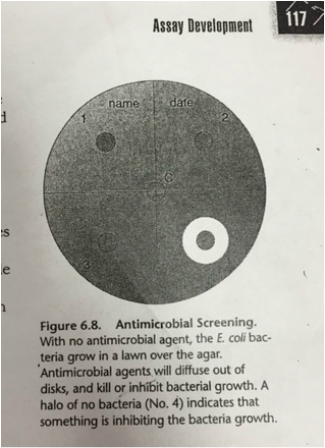
13.) You should have 6 Petri plates, each containing a negative control in the middle, a positive control, and 3 sample disks (picture of figure 6.8 from worksheet below). Record exactly which plant extracts with which solvent went into each quadrant. Let everything "soak in" for a few minutes.
9.) Transfer 1 mL of E. coli broth to the middle of each Petri dish using a sterile pipet. Sterilize a spreading loop (using alcohol and flame), and evenly spread the bacterial culture around Petri dish. Once finished, quickly cover and let the culture soak into the agar for about 15 minutes.
10.) Carefully place 1 disk into the middle of each quadrant with about 2 cm from the outer edge of the Petri dish using sterile forceps. Dry any left over liquid before placing the disk on the Petri dish. Make sure all the methanol-extracted samples are on the same dish and all the water-extracted samples are on the same dish.
11.) Repeat Step 10 2 more times so that you have 3 copies of methanol extractions and 3 copies of deionized water extractions.
12.) Place 1 of the negative control disks, of either sterile distilled water or methanol, in the center of the appropriate plate. Place 1 positive control disk with ampicillin in another quadrant of each plate.
13.) You should have 6 Petri plates, each containing a negative control in the middle, a positive control, and 3 sample disks (picture of figure 6.8 from worksheet below). Record exactly which plant extracts with which solvent went into each quadrant. Let everything "soak in" for a few minutes.
|
14.) Be sure the disks are attached well to the surface of the agar. Invert the plates and incubate at 37*C for 24-48 hours.
15.) After the incubation period, observe the plates with the plant extract disks for zones pf inhibition. This is a clear area formed around disk by the inhibitory action of a substance(s) in the plant material. Take a picture or draw your plates, labeling any inhibition of bacterial growth. 16.) Make a data table of your results. Describe the bacterial lawn around each disk. Then measure and record the diameter and clarity of any cleared areas around the disks. Make sure to give measurements of observations. |
|
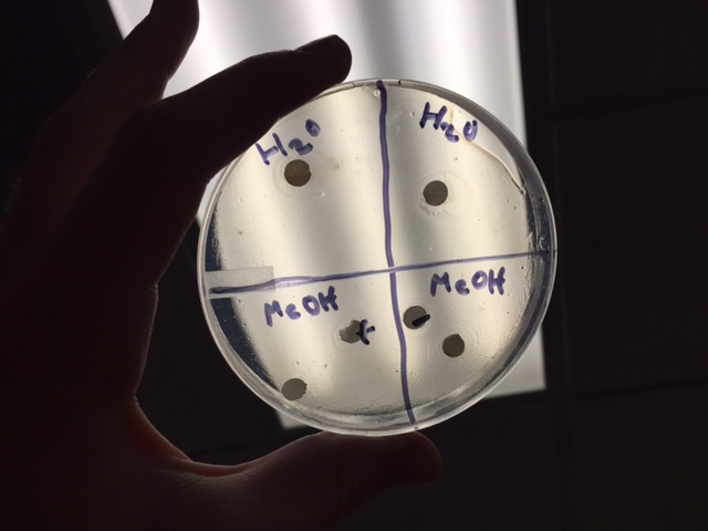
~Results:
The results of my were a little mixed up. I accidentally labeled the methanol incorrectly (switching the negative/positive), but the results were pretty much fine. The H₂O, however, were not the same, as they were suppose to be. One is negative and the other is positive. I don't honestly know why they weren't the same. I wasn't at school on Monday so I missed a lot of the explanation of this lab :( |
|
~Data Analysis/Conclusion:
It's hard to say what I did when I didn't quite understand what I was doing. I partnered up with Rick when I came back to school on Tuesday and did pretty much what he was doing. I probably should have asked more than just "Am I doing this correctly?" Labs are so hard for me for some odd reason and it annoys the living day lights out of me. Guess I could talk to my teacher about this later. Probably should.
It's hard to say what I did when I didn't quite understand what I was doing. I partnered up with Rick when I came back to school on Tuesday and did pretty much what he was doing. I probably should have asked more than just "Am I doing this correctly?" Labs are so hard for me for some odd reason and it annoys the living day lights out of me. Guess I could talk to my teacher about this later. Probably should.
